March 9, 2023
Corey Montgomery, M.D. Among Pioneers Recognized in Black History Wall of Honor Exhibit
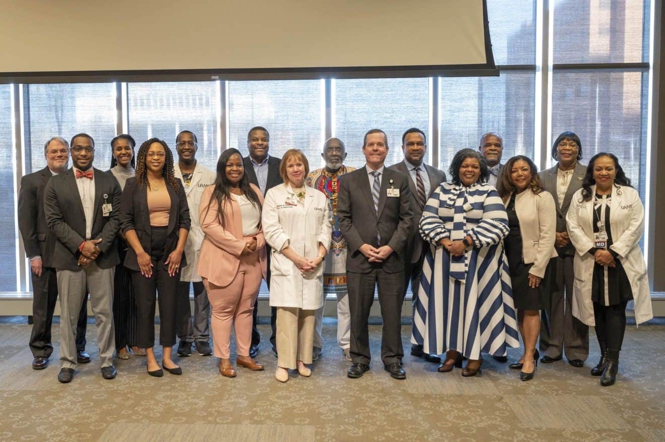
The University of Arkansas for Medical Sciences (UAMS) Division for Diversity, Equity and Inclusion (DDEI) held a Feb. 28 ceremony to recognize members of UAMS’ 2023 Black History Wall of Honor Exhibit.
The exhibit, which was displayed in the hospital’s first-floor admissions and discharge area during Black History Month, highlighted the accomplishments of Black pioneers from UAMS’ past and present. Many of the honorees attended the closing ceremony.
Cam Patterson, M.D., MBA, UAMS chancellor and CEO of UAMS Health, praised the honorees and thanked them for their efforts to improve health care in Arkansas.
“UAMS would not be what it is without these individuals who made so much of their time here,” he said. “This exhibit is so powerful, and it says so much about our values and our commitment to equity and social justice.”
Tim Nutt, director of the UAMS Historical Research Center, said the exhibit showed the rich history of UAMS and the significant accomplishments of its pioneers. He also acknowledged the challenges many of them faced, overcoming racism, sexism and elitism as they blazed trails for others to follow.
Nutt highlighted the stories of two honorees, Edith Irby Jones, M.D., and Joycelyn Elders, M.D. In 1948, Jones was the first African American admitted to the UAMS College of Medicine. She graduated in 1952 and became a renowned physician who inspired others to pursue medical careers. One of those was Elders, who graduated from UAMS in 1960 and went on to serve as the United States’ first Black surgeon general from 1993-94.
“UAMS is not the same institution it was in 1948 when Dr. Jones applied,” Nutt said. “In the past 75 years, long strides have been made toward diversity, equity and inclusion across the institution, and progress continues to be seen every day.”
Michelle Krause, M.D., MPH, the CEO of UAMS Medical Center and senior vice chancellor for UAMS Health, said diversity is a vital component in the work of UAMS’ faculty and staff. That diversity made a difference in the early days of the COVID-19 pandemic, she said, as UAMS was able to reach individuals and communities from a variety of backgrounds and provide the level of care they needed.
“We’re dedicated to closing the gaps where we have health care disparities, both at UAMS and across our state,” she said.
Hosea Long, an honoree and former associate vice chancellor for human resources, said that after he arrived at UAMS in 1991, he became part of a group that pushed to formalize diversity and inclusion efforts on the campus. He said UAMS has accomplished that goal in part by creating a leadership position dedicated to diversity, equity and inclusion. That role is held by Vice Chancellor Brian Gittens, Ed.D., MPA.
“With UAMS being the only academic health institution in the state, it should be a proponent of diversity and inclusion,” said Long, who retired in 2013 after more than 20 years at UAMS. “I see that happening here, and it’s very exciting.”
The 2023 Black History Wall of Honor Exhibit was created through a partnership involving DDEI, the UAMS Historical Research Center and the Wall of Honor Planning Committee.
Betholyn Gentry, Ph.D., who retired in 2020 from her role as a speech-language pathology professor in the College of Health Professions, said she appreciated being recognized in the exhibit. Gentry was the first Black faculty member in the speech-language pathology program, where she served for 43 years.
“I’ll be glad when we get to the day when there are no more ‘first’ African Americans in any position, when it’s just a normal part of the hiring process,” she said. “We’re getting there. The people being honored today are a testament to UAMS’ attempts to foster diversity.”
Lanita White, Pharm.D., CEO of Community Health Centers of Arkansas and former assistant dean for student affairs in the UAMS College of Pharmacy, said she felt honored to be included in the exhibit, accompanying colleagues whom she described as “health care giants.”
“For UAMS to collectively pause and honor these folks for their accomplishments, it speaks to a continuing focus on diversity, equity and inclusion,” she said. “We’re making sure that focus is front and center not only for our staff and students but also for our patients.”
The members of the 2023 Black History Wall of Honor Exhibit are:
Bill Bauknight
Keneshia Bryant-Moore, Ph.D., RN
Dana Carthon, Ph.D., RN
Ashley Connors, BSN, RN
Rodney Davis, M.D.
Joycelyn Elders, M.D.
Pebbles Fagan, Ph.D., MPH
Henry Foster, M.D.
Betholyn Gentry, Ph.D.
Brian Gittens, Ed.D., MPA
Sharon D. Harris, Pharm.D.
Ronda Henry-Tillman, M.D.
Barbara Johnson, MSN, RN
Patricia J. Johnson
Dina M. Jones, Ph.D., MPH
Edith Irby Jones, M.D.
Roy Kitchen, MBA
Hosea Long
Patricia Marks, Ph.D.
Freda McKissic Bush
Kevin Means, M.D.
Brooke E.E. Montgomery, Ph.D., MPH
Corey Montgomery, M.D.
Lenora S. Newsome, Pharm.D.
Austin Porter, DrPH, MPH
Phillip Leon Rayford, Ph.D.
Al Reece, M.D.
Gloria Richard-Davis, M.D., MBA
Ray Smith, RRT
Billy Thomas, M.D.
Charles White
Lanita White, Pharm.D.
Sophronia Reacie Williams, RN
Sterling Williams, M.D., Ph.D.
Olivia Wilson, MS
The closing ceremony also marked the conclusion of DDEI’s commemorations for Black History Month. On Feb. 27, DDEI hosted a virtual panel discussion — titled “Resist Silence: Lift Every Voice!” — that focused on ways to empower diverse voices and opinions.
Gloria Richard-Davis, M.D., MBA, executive director of DDEI, moderated the discussion, which included topics such as creating safe spaces, the stigmas regarding mental health in the Black community, the importance of mentoring, why allies matter, and how one’s faith can empower one’s voice. The panelists were Mildred Randolph, DVM, director of the UAMS Seeking Educational Equity and Diversity (SEED) program; Tyrun Haynie, emergency preparedness director for Institutional Support Services; Isis Pettway, M.S., licensed associate counselor with the UAMS Health AR ConnectNow program in the Psychiatric Research Institute; Bill Ventres, M.D., associate professor in the College of Medicine; and Keneshia Bryant-Moore, Ph.D., RN, associate professor in the College of Public Health.